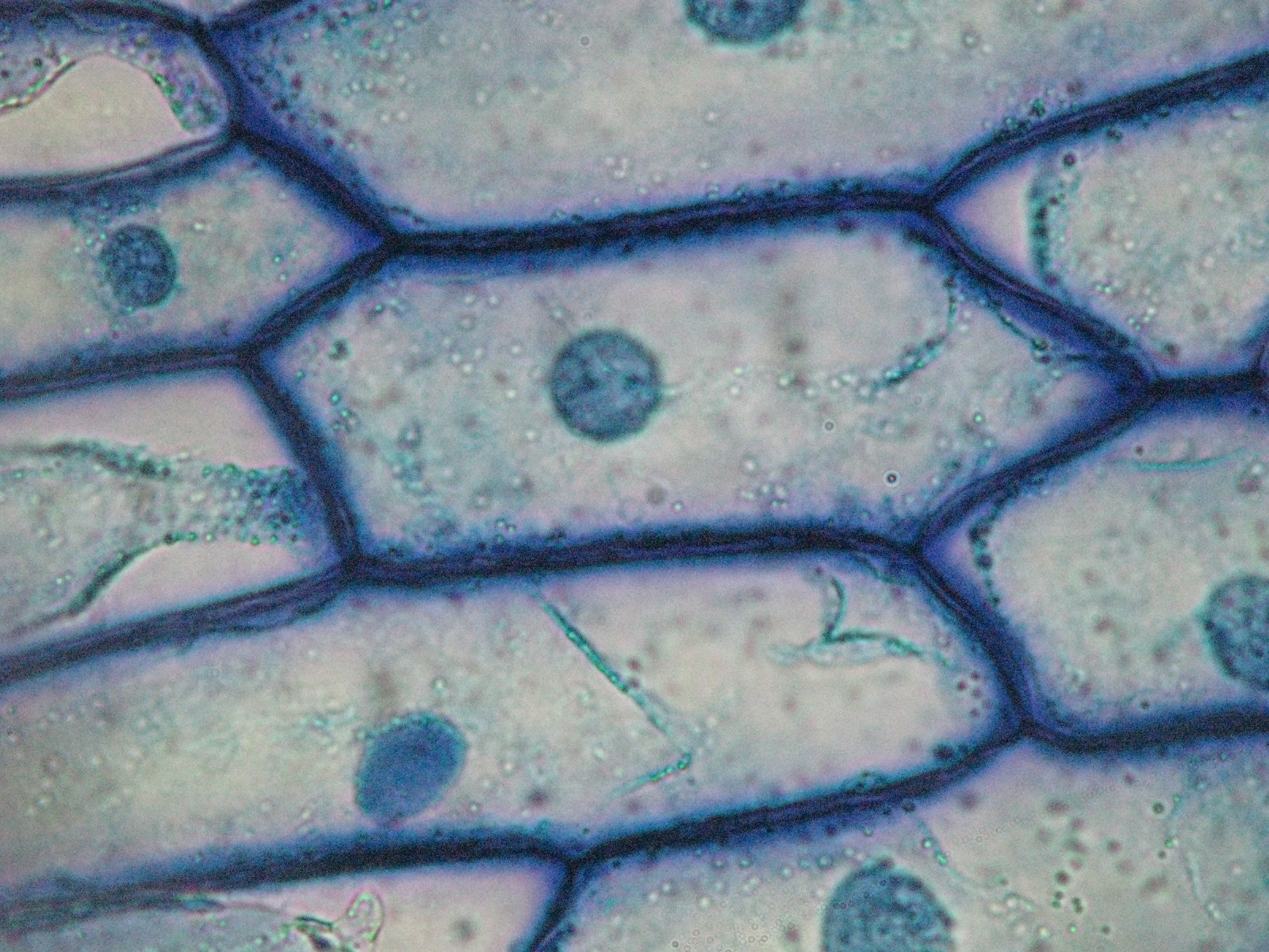
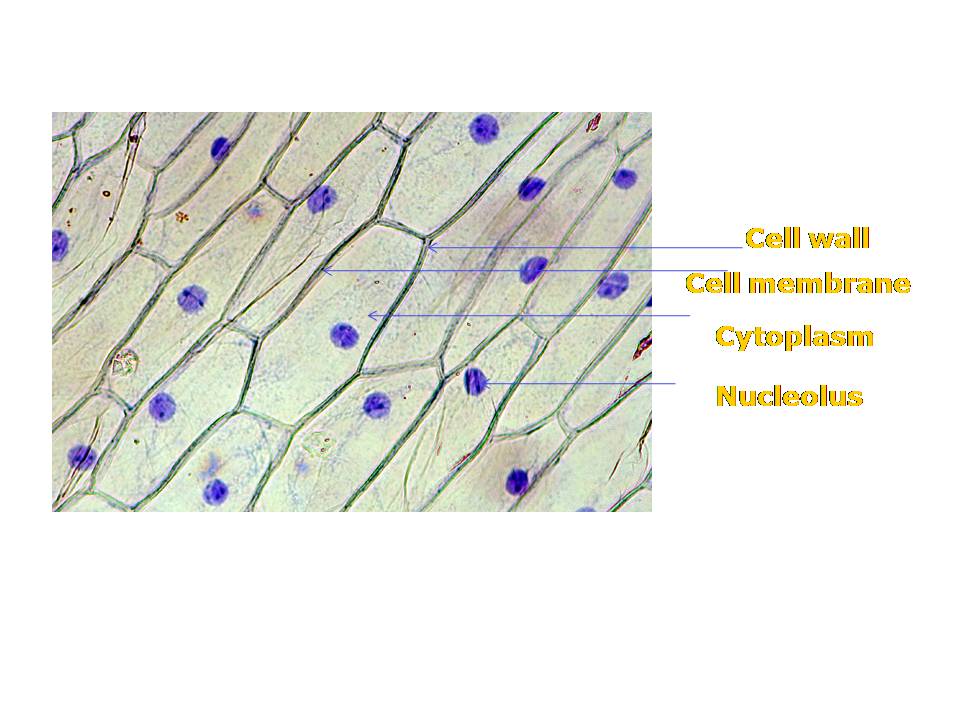
Onion Skin Cell Diagram
Onion epidermal cell diagram Onion cell cells microscope micrograph microscopic stock skin alamy magnification root tip section allium high epidermis bulb mitosis photomicrograph show Lm of onion skin
Onion Cells under Microscope
Cells onion cell microscope under iodine epidermis plant bulb 10x mount inner skin light bubbles air animal nucleus eukaryotic microscopy Onion cells microscope hi-res stock photography and images Onion_cells – biobiznews
Biopedia: practicals
Onion cell microscope hi-res stock photography and imagesOnion mount temporary cells labelled draw cell prepare peel diagrams blissful earth objective observations record Cells cell plant vacuole onion microscope under structure microscopic do wall peel nucleus biology lab membrane diagram epidermal identify slideOnion cells red file size wikipedia.
Blissful earthOnion cells under microscope Onion microscope magnified 40x 100x microscopyRevision notes for science chapter 8.

Onion cell cells microscope micrograph microscopic alamy stock skin magnification section high cepa allium mitosis root x100
Onion skin lmImage gallery onion cell Can someone please post me a diagram ofOnion cell epidermal peel size.
Onion scienceOnion cells beautiful world Onion cell peel draw cytoplasm membrane vacuole showing brainly figureBeautiful world: onion cells.
![Can someone please post me a diagram of - 1] Onion cells 2] Animal](https://1.bp.blogspot.com/_ATZV16R8qIg/TI251c78pQI/AAAAAAAAAB8/j9iX_9Voagw/s320/onion.jpeg)
Onion biology uses
Onion epidermal drawings epidermis labeled biology chromosomes chromosome dna observationFun science blog: onion cell File:red onion cells.jpgDraw the figure of an onion peel showing cell.
.


Beautiful World: Onion cells
File:Red Onion Cells.JPG - Wikipedia

Biopedia: Practicals

draw the figure of an onion peel showing cell - Brainly.in

Onion cells microscope hi-res stock photography and images - Alamy

Onion Cells under Microscope

LM of Onion Skin - Stock Image - C012/1141 - Science Photo Library
Fun Science Blog: Onion Cell

Revision Notes for Science Chapter 8 - Cell — Structure and functions