Onion Cell Diagram
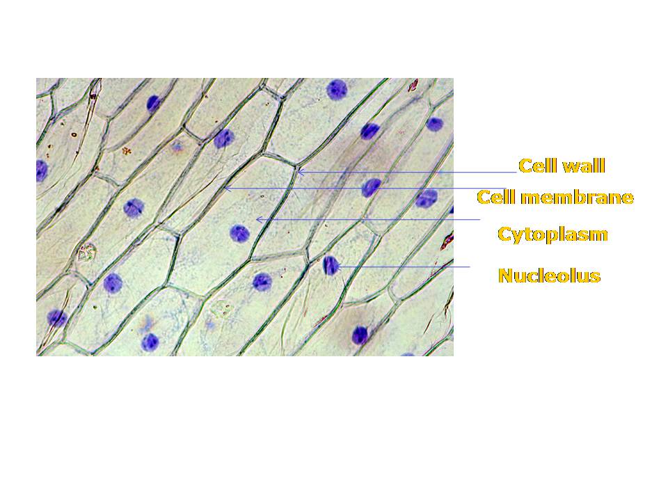
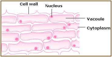
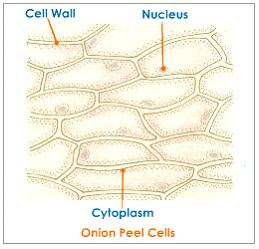
Onion cell diagram drawing Onion cells under microscope Onion cell cells microscope micrograph microscopic stock skin alamy magnification root tip section allium high epidermis bulb mitosis photomicrograph show
Blissful Earth
Onion cell epidermal peel size Blissful earth Onion cell microscope hi-res stock photography and images
Lab slides. cell types
Onion cells cell lab types slidesOnion cell diagram drawing Microscope epidermis 400x membrane onionsOnion mount temporary cells labelled draw cell prepare peel diagrams blissful earth objective observations record.
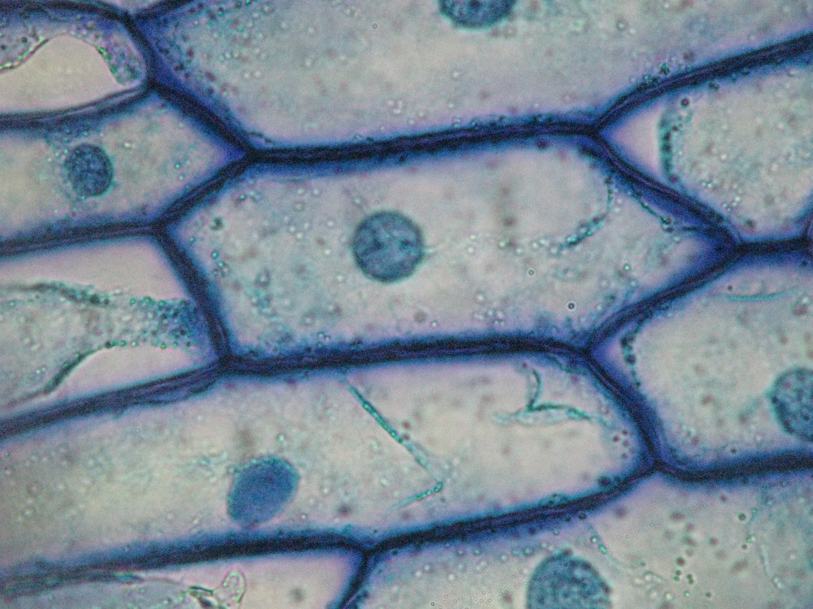
Onion cellsOnion cell cells diagram structure 2010 biology microscopic occupies september uses introduction Cebola zwiebel cipolla cells micrografia micrografo mikrograph microscopio pinoOnion skin under microscope 400x.

Onion cells 2
Magnified microscope cell 40x microscopy micrographs wallsCell peel ncert Onion cell 400x lab microscope under labeled cells structure scoop science white lookedThe science scoop: onion cell lab.
Onion epidermal drawings epidermis labeled biology chromosomes chromosome dna observationOnion cells beautiful world Onion microscope magnified 40x 100x microscopyBiopedia: practicals.

Onion cells
Beautiful world: onion cellsOnion cell diagram drawing Onion epidermal cell diagramBiology help online: september 2010.
Microscope methylene cells labelled typical epidermal biologicalOnion cells under microscope .


Onion Cell Diagram Drawing - lana1970

Onion Cell Diagram Drawing - lana1970

Blissful Earth

Beautiful World: Onion cells

Onion Cells under Microscope

Onion cells 2 | Basically the same of onion cells 1 but this… | Flickr
.PNG)
Lab Slides. Cell Types - Presentation Biology

Biology help online: September 2010

Onion cells | High-Quality Nature Stock Photos ~ Creative Market